Fetal Skull Superior View: A Detailed Guide
Services
Welcome to Unilevel Studios' detailed exploration of the superior view of the fetal skull. In this comprehensive guide, we delve into the intricate anatomy and unique features of the fetal skull when viewed from the top.
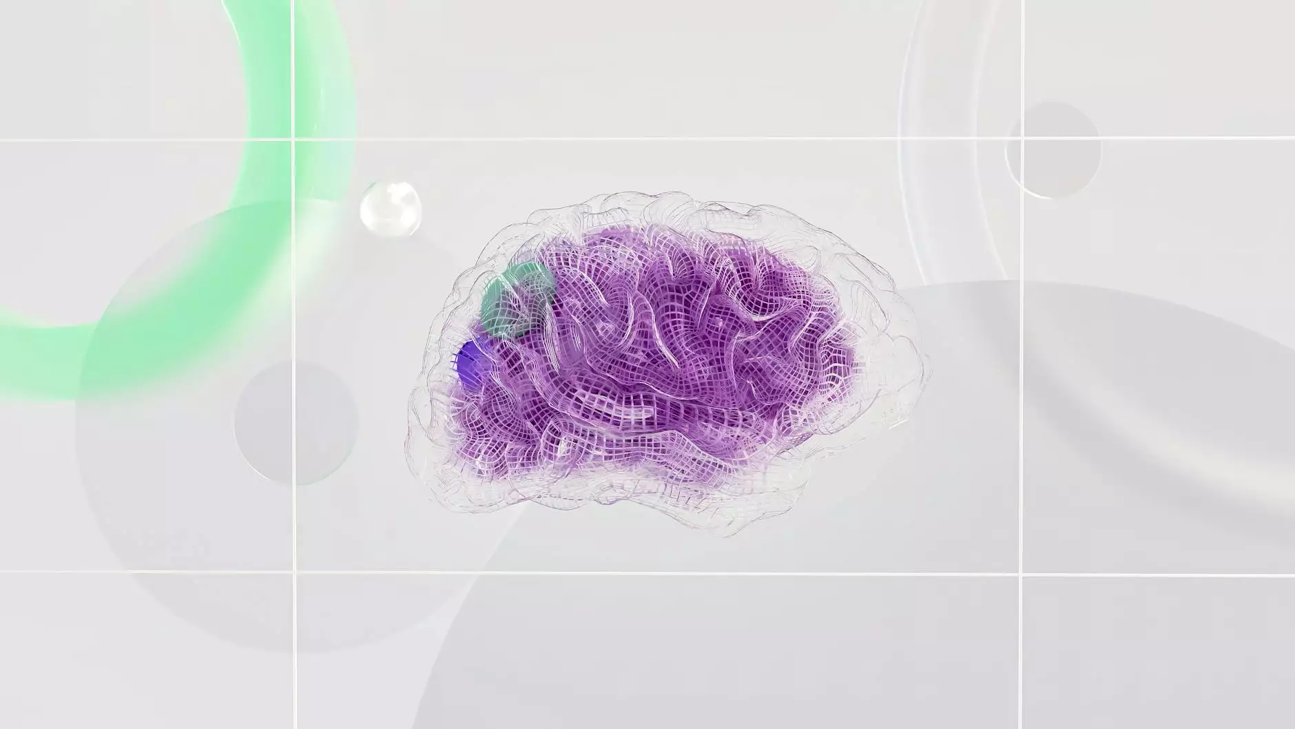
The Anatomy of the Fetal Skull
The fetal skull is a remarkable structure that undergoes numerous transformations during the developmental process in the womb. When viewed from the top, also known as the superior view, several key features become evident.
Fontanelles and Sutures
One of the defining characteristics of the fetal skull in the superior view is the presence of fontanelles and sutures. Fontanelles, also referred to as soft spots, are areas where the bones of the skull have not yet fused together, allowing for flexibility during childbirth. The sutures are the joints between the skull bones that facilitate the growth of the skull.
Cranial Bones
The fetal skull comprises several cranial bones that come together to form a protective enclosure for the developing brain. These bones include the frontal bone, parietal bones, temporal bones, occipital bone, sphenoid bone, and ethmoid bone.
Developmental Stages
As the fetus grows and develops, the fetal skull undergoes a series of remarkable changes. Understanding these developmental stages is essential for healthcare providers, anatomists, and researchers studying fetal development.
Early Gestation
During early gestation, the fetal skull is primarily composed of cartilage, which gradually ossifies to form the bony structures seen in the mature skull. At this stage, the fontanelles are prominent, allowing for easier passage through the birth canal.
Mid to Late Gestation
As the fetus continues to develop, the cranial bones become more defined, and the sutures between them become visible. This stage marks significant growth and maturation of the fetal skull, preparing the fetus for birth.
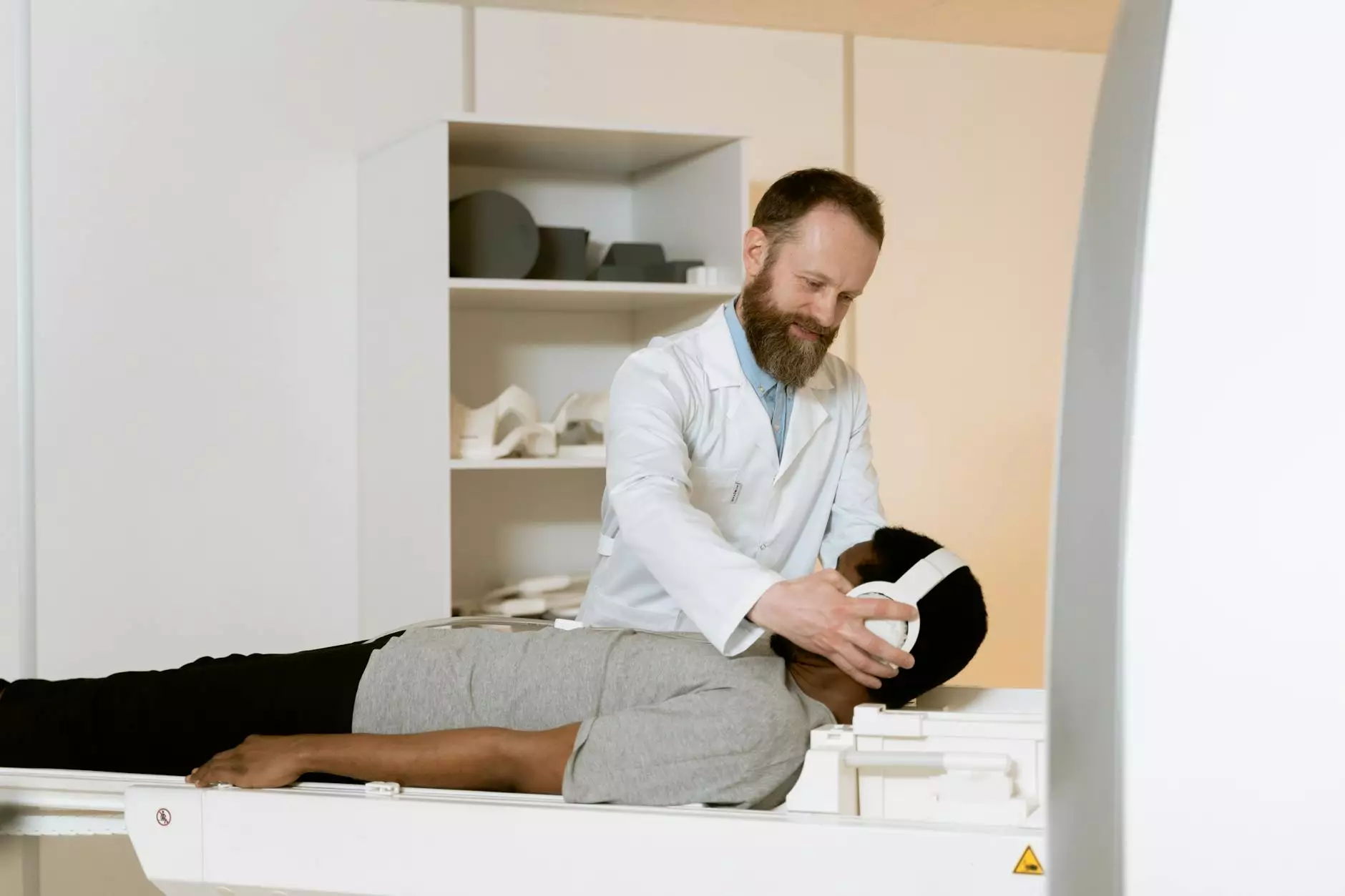
Clinical Considerations
Knowledge of the fetal skull's superior view is crucial in various clinical settings, including obstetrics, radiology, and pediatrics. Understanding the normal anatomy and potential variations in the fetal skull can aid in the diagnosis and management of congenital abnormalities.
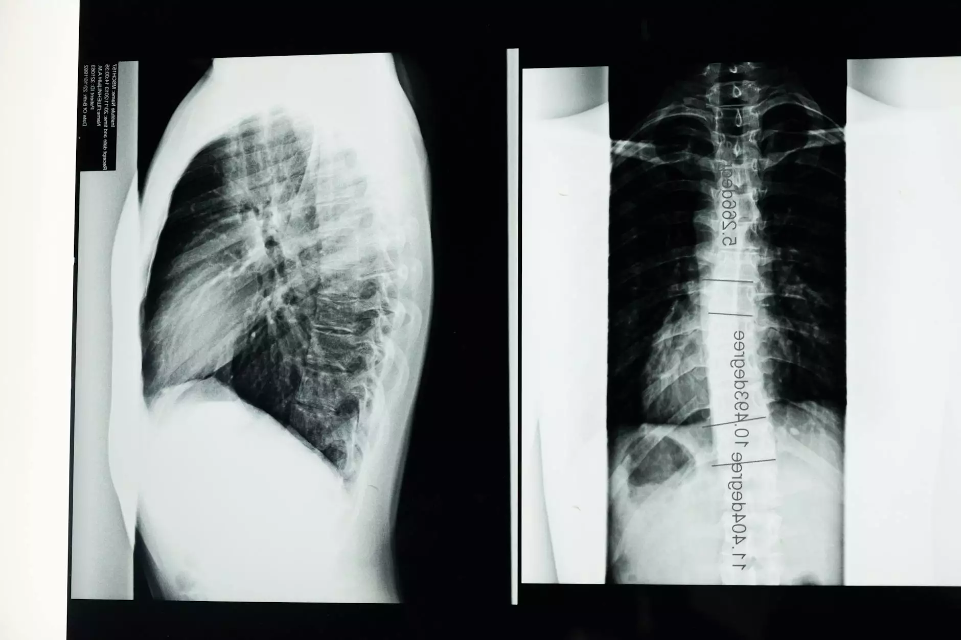
Ultrasound Imaging
Ultrasound technology has revolutionized the visualization of the fetal skull during pregnancy. Obstetric ultrasonography allows healthcare providers to assess the fetal skull's development, detect anomalies, and monitor growth with high precision.
Pathological Conditions
Certain pathological conditions can affect the development of the fetal skull, leading to structural abnormalities or deformities. These conditions may require specialized medical care and interventions to ensure the health and well-being of the fetus.
Conclusion
In conclusion, exploring the superior view of the fetal skull offers valuable insights into the intricate world of fetal development. At Unilevel Studios, we are dedicated to providing informative content on a wide range of topics, including the anatomy of the fetal skull.
Stay tuned for more educational resources and expert knowledge from Unilevel Studios, your trusted partner in website development and business services.